ALICA
Automated Laser Illumination Control Algorithm
About
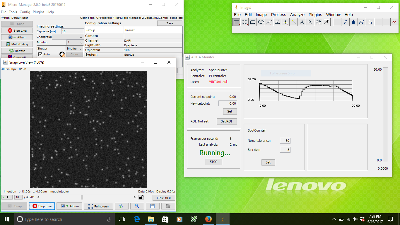
ALICA is an open-source Micro-Manager plugin for real-time control of single molecule photodynamics using adaptive illumination. In particular, ALICA enables autonomous super-resolution fluorescence imaging using techniques such as STORM and PALM [1] [2] [3].
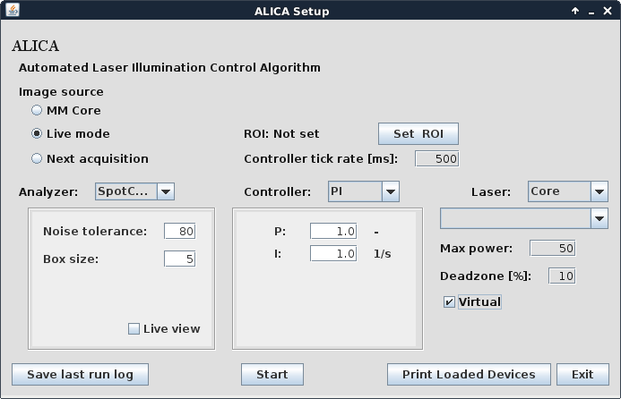
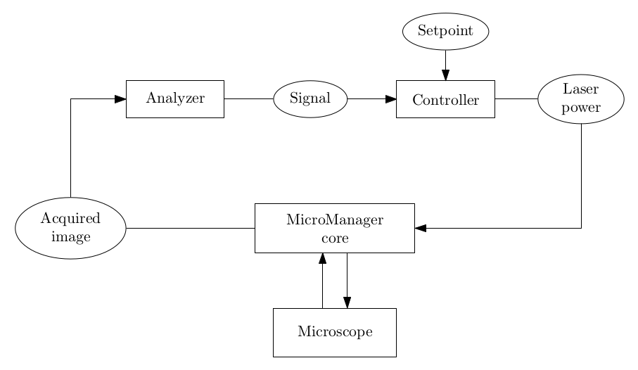
ALICA works by analyzing the incoming images during an acquisition to produce an estimate of the number of fluorescence emitting molecules in a region of interest. The estimates are then fed into a control system that automatically adjusts the illumination intensity to maintain the optimum density of emitting molecules for the desired application. Example applications include
- STORM and PALM super-resolution fluorescence microscopy
- in vitro single molecule assays
- single particle tracking
A primary design goal of ALICA is extensibility. A minimal knowledge of the Java programming language will allow you to write your own analyzers for deriving quantitative information from an image stream and controllers for implementing closed-loop feedback for your hardware. As a Micro-Manager plugin, ALICA easily integrates with many types of cameras and illumination sources.
ALICA was designed and written by Marcel Stefko and Kyle M. Douglass in the Laboratory of Experimental Biophysics at the EPFL to automate the lab’s STORM and PALM microscopes.
Acknowledgments
Thanks
- The Laboratory of Experimental Biophysics and Suliana Manley
- The École Polytechnique Fédérale de Lausanne
- Micro-Manager and the Micro-Manager community
- AutoLase by Seamus Holden and Thomas Pengo
- QuickPALM by Ricardo Henriques, et al.
- SpotCounter by Nico Stuurman
Authors
See Also
- SASS - STORM Acquisition Simulation Software
Footnotes
| [1] | M. J. Rust, M. Bates, and X. Zhuang, “Sub-diffraction-limit imaging by stochastic optical reconstruction microscopy (STORM)”, Nature Methods 3, 793-796 (2006). http://www.nature.com/nmeth/journal/v3/n10/abs/nmeth929.html |
| [2] | E. Betzig, et al., “Imaging intracellular fluorescent proteins at nanometer resolution,” Science 313, 1642-1645 (2006). http://science.sciencemag.org/content/313/5793/1642 |
| [3] | S. T. Hess, T. P. K. Girirajan, and M. D. Mason, “Ultra-high resolution imaging by fluorescence photoactivation localization microscopy,” Biophysical Journal 91, 4258-4272 (2006). http://www.sciencedirect.com/science/article/pii/S0006349506721403 |